Copolymer dynamics


M. Zakaria Slimani, A.J. Moreno, G. Rossi, J. Colmenero. Dynamic Heterogeneity in Random and Gradient Copolymers: A Computational Investigation. Macromolecules, ASAP, 2013. DOI: 10.1021/ma400577d

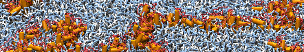
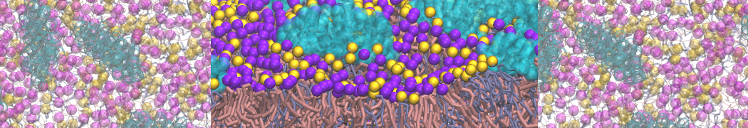
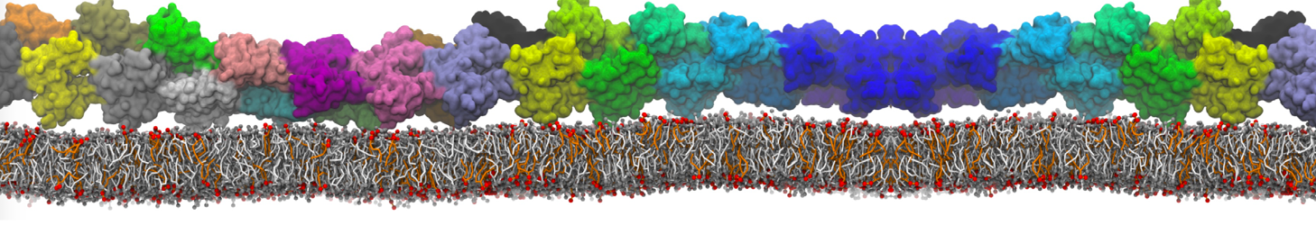
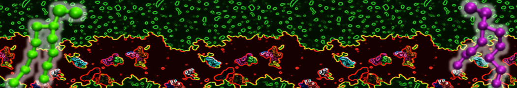
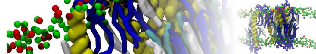
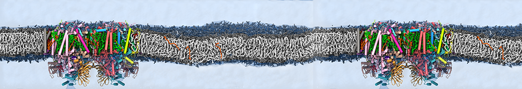
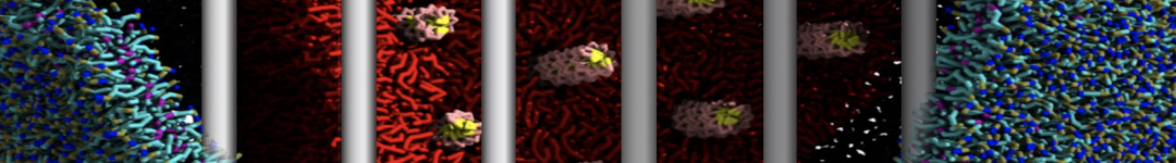
By means of molecular dynamics simulations, we investigate the structural relaxation in disordered random copolymers and lamellar phases of gradient copolymers, containing chemical species of very different mobilities. Two models have been investigated: a generic bead–spring system and a MARTINI coarse-grained model of a polyester resin. The lamellar phase of the gradient copolymer is formed by domains rich in one species and poor in the other one, which are separated by broad interfaces. Unlike in strongly segregated block copolymers, there is a finite probability of finding monomers of a given species at any position within the domains rich in the other species. A direct consequence of this feature is that monomers can probe very different chemical environments, and because of the strong dynamic asymmetry between the two components, their relaxation are characterized by an extreme dynamic heterogeneity. This is confirmed by an analysis of dynamic correlators as a function of the distance to the interface. In the case of random copolymers long-range ordering is not possible, and local microsegregation results in a much weaker dynamic heterogeneity. The former features are consistent with the experimental observation of narrow glass transitions in random copolymers but extremely broad ones in lamellar gradient copolymers.

folding lipid monolayers

"Folding of lipid monolayers containing lung surfactant proteins SP-B1–25 and SP-C studied via coarse-grained molecular dynamics simulations" by S.L. Duncan, R.G. Larson. Biochimica et Biophysica Acta - Biomembranes, 2010, 1798, 1632-1650

To explore the role of lung surfactant proteins SP-B and SP-C in storing and redelivering lipid from lipid monolayers during the compression and re-expansion occurring in lungs during breathing, we simulate the folding of lipid monolayers with and without these proteins. We utilize the MARTINI coarse-grained force field to simulate monolayers containing pure dipalmitoylphosphatidylcholine (DPPC) and DPPC mixed with palmitoyloleoylphosphatidylglycerol (POPG), palmitic acid (PA), and/or peptides. The peptides considered include the 25-residue N-terminal fragment of SP-B (SP-B125), SP-C, and several SP-B125 mutants in which charged and hydrophilic residues are replaced by hydrophobic ones, or vice-versa. We observe two folding mechanisms: folding by the amplification of undulations and folding by nucleation about a defect. The first mechanism is observed in monolayers containing either POPG or peptides, while the second mechanism is observed only with peptides present, and involves the lipid-mediated aggregation of the peptides into a defect, from which the fold can nucleate. Fold nucleation from a defect displays a dependence on the hydrophobic character of the peptides; if the number of hydrophobic residues is decreased significantly, monolayer folding does not occur. The addition of POPG or peptides to the DPPC monolayer has a fluidizing effect, which assists monolayer folding. In contrast, the addition of PA has a charge-dependent condensing affect on DPPC monolayers containing SP-C. The peptides appear to play a significant role in the folding process, and provide a larger driving force for folding than POPG. In addition to promoting fold formation, the peptides also display fusogenic behavior, which can lead to surface refining.

Cardiolipin- cyctochrome binding

C. Arnarez, J.P. Mazat, J. Elezgaray, S.J. Marrink, X. Periole. Evidence for cardiolipin binding sites on the membrane-exposed surface of the cytochrome bc1. JACS, in press, 2013. DOI: 10.1021/ja310577u. abstract


and

C. Arnarez, S.J. Marrink, X. Periole. Identification of cardiolipin binding sites on cytochrome c oxidase at the entrance of proton channels. Sci. Rep., 3:1263, 2013. open access

Two papers revealing cardiolipin binding sites on the surface of membrane embedded respiratory chain complexes III and IV.

solvent-exposior

"Solvent-Exposed Tails as Prestalk Transition States for Membrane Fusion at Low Hydration" by Y.G. Smirnova, S.J. Marrink, R. Lipowsky and V. Knecht. J. Am. Chem. Soc., 2010, 132, 6710–6718

Membrane fusion is a key step in intracellular trafficking and viral infection. The underlying molecular mechanism is poorly understood. We have used molecular dynamics simulations in conjunction with a coarse grained model to study early metastable and transition states during the fusion of two planar palmitoyl-oleoyl-phosphatidylcholine (POPC) bilayers separated by five waters per lipid in the cis leaflets at zero tension. This system mimics the contact area between two vesicles with large diameters compared to the membrane thickness at conditions where fusion may start in the core of the contact area. At elevated temperatures, the two proximal leaflets become connected via multiple lipid molecules and form a stalklike structure. At room temperature, this structure has a free energy of 3kBT and is separated from the unconnected state by a significant free energy barrier of 20kBT. Stalk formation is initiated by the establishment of a localized hydrophobic contact between the bilayers. This contact is either formed by two partially splayed lipids or a single fully splayed one leading to the formation of a (metastable) splayed lipid bond intermediate. These findings indicate that, for low hydration, early membrane fusion kinetics is not determined by the stalk energy but by the energy of prestalk transition states involving solvent-exposed lipid tails.

RNA official release

Always dreamt about mixing RNA with other Martini ingredients? Here is your chance: RNA official release.

cyclic peptides

"On the Antibacterial Action of Cyclic Peptides: Insights from Coarse-Grained MD Simulations" by A. Khalfa and M. Tarek. J. Phys. Chem. B, 2010, 114, 2676–2684.

[RRKWLWLW] cyclic peptides have been shown to exhibit remarkable in vitro and in vivo antibacterial activity. Peptides alike seem to be promising for the development of new compounds to combat microbial pathogens, yet the molecular level understanding of their mechanism of action remains unclear. Here, we use coarse-grained (CG) molecular dynamics (MD) simulations of these cyclic peptides interacting with antibacterial cytoplasmic membrane models composed of a mixture of palmitoyl-oleoyl-phosphatidyl-ethanolamine (POPE) and palmitoyl-oleoyl-phosphatidylglycerol (POPG) lipid bilayers to provide a better understanding of their mode of action. In particular, the MD simulations performed at various concentrations of membrane-bound cyclic peptides reveal a novel type of mechanism in which the peptides first self-assemble at the membrane interface into amphipathic nanotubes. At high enough concentrations, coating of the membrane causes extrusion of lipids from the exposed bilayer leaflet, leading ultimately to a release of phospholipid micellar aggregates. Furthermore, the cyclic peptides also induce a drastic change in the lateral pressure profiles of the exposed leaflet, indicating a direct effect on the mechanical properties of the bilayer.