Article22

"Lung Surfactant Protein SP-B Promotes Formation of Bilayer Reservoirs from Monolayer and Lipid Transfer between the Interface and Subphase", S. Baoukina and D.P. Tieleman, Biophys. J., 100:1678-1687, 2011

We investigated the possible role of SP-B proteins in the function of lung surfactant. To this end, lipid monolayers at the air/water interface, bilayers in water, and transformations between them in the presence of SP-B were simulated. The proteins attached bilayers to monolayers, providing close proximity of the reservoirs with the interface. In the attached aggregates, SP-B mediated establishment of the lipid-lined connection similar to the hemifusion stalk. Via this connection, a lipid flow was initiated between the monolayer at the interface and the bilayer in water in a surface-tension-dependent manner. On interface expansion, the flow of lipids to the monolayer restored the surface tension to the equilibrium spreading value. SP-B induced formation of bilayer folds from the monolayer at positive surface tensions below the equilibrium. In the absence of proteins, lipid monolayers were stable at these conditions. Fold nucleation was initiated by SP-B from the liquid-expanded monolayer phase by local bending, and the proteins lined the curved perimeter of the growing fold. No effect on the liquid-condensed phase was observed. Covalently linked dimers resulted in faster kinetics for monolayer folding. The simulation results are in line with existing hypotheses on SP-B activity in lung surfactant and explain its molecular mechanism.

Article21


"Controlled Self-Assembly of Filled Micelles on Nanotubes", N. Patra and P. Kral, J. Am. Chem. Soc., 2011 (Communication) DOI: 10.1021/ja2009778


We have used coarse-grained molecular dynamics simulations to show that hydrated lipid micelles of preferred sizes and amounts of filling with hydrophobic molecules can be self-assembled on the surfaces of carbon nanotubes. We simulated micelle formation on a hydrated (40,0) carbon nanotube with an open end that was covered with amphiphilic double-headed CH3(CH2)14CH(((CH2OCH2CH2)2(CH2COCH2))2H)2 or single-headed CH3(CH2)14CH2((CH2OCH2CH2)2(CH2COCH2))4H lipids and filled with hexadecane molecules. Once the hexadecane molecules inside the nanotube were pressurized and the lipids on its surface were dragged by the water flowing around it, kinetically stable micelles filled with hexadecane molecules were sequentially formed at the nanotube tip. We investigated the stability of the thus-formed kinetically stable filled micelles and compared them with thermodynamically stable filled micelles that were self-assembled in the solution.

Article20


"Caught in the Act: Visualization of SNARE-Mediated Fusion Events in Molecular Detail" H. Jelger Risselada, Carsten Kutzner and Helmut Grubmüller, ChemBioChem, 12, 2011.

Neurotransmitter release at the synapse requires fusion of synaptic vesicles with the presynaptic plasma membrane. SNAREs are the core constituents of the protein machinery responsible for this membrane fusion, but the actual fusion mechanism remains unclear. Here, we have simulated neuronal SNARE-mediated membrane fusion in molecular detail. In our simulations, membrane fusion progresses through an inverted micelle fusion intermediate before reaching the hemifused state. We show that at least one single SNARE complex is required for fusion, as has also been confirmed in a recent in vitro single-molecule fluoresence study. Further, the transmembrane regions of the SNAREs were found to play a vital role in the initiation of fusion by causing distortions of the lipid packing of the outer membrane leaflets, and the C termini of the transmembrane regions are associated with the formation of the fusion pores. The inherent mechanical stress in the linker region of the SNARE complex was found to drive both the subsequent formation and expansion of fusion pores. Our simulations also revealed that the presence of homodimerizations between the transmembrane regions leads to the formation of unstable fusion intermediates that are under high curvature stress. We show that multiple SNARE complexes mediate membrane fusion in a cooperative and synchronized process. Finally, we show that after fusion, the zipping of the SNAREs extends into the membrane region, in agreement with the recently resolved X-ray structure of the fully assembled state.

Article19


"Inhibition of peptide aggregation by lipids: Insights from coarse-grained molecular simulations" A. Hung and I.P.P. Yarovsky, J. Mol. Graph. Mod., 29:597-607, 2011.

The amyloidogenic peptide apolipoprotein C-II(60-70) is known to exhibit lipid-dependent aggregation behaviour. While the peptide rapidly forms amyloid fibrils in solution, fibrillisation is completely inhibited in the presence of lipids. In order to obtain molecular-level insights into the mechanism of lipid-dependent fibril inhibition, we have employed molecular dynamics simulations in conjunction with a coarse-grained model to study the aggregation of an amyloidogenic peptide, apoC-II(60-70), in the absence and presence of a short-chained lipid, dihexanoylphosphatidylcholine (DHPC). Simulation of a solution of initially dispersed peptides predicts the rapid formation of an elongated aggregate with an internal hydrophobic core, while charged sidechains and termini are solvent-exposed. Inter-peptide interactions between aromatic residues serve as the principal driving force for aggregation. In contrast, simulation of a mixed peptide-DHPC solution predicts markedly reduced peptide aggregation kinetics, with subsequent formation of a suspension of aggregates composed of smaller peptide oligomers partially inserted into lipid micelles. Both effects are caused by strong interactions between the aromatic residues of the peptide with the lipid hydrophobic tails. This suggests that lipid-induced aggregate inhibition is partly due to the preferential binding of peptide aromatic sidechains with lipid hydrophobic tails, reducing inter-peptide hydrophobic interactions. Furthermore, our simulations suggest that the morphology of peptide aggregates is strongly dependent on their local lipid environment, with greater contacts with lipids resulting in the formation of more elongated aggregates. Finally, we find that peptides disrupt lipid self-assembly, which has possible implications for explaining the cytotoxicity of peptide oligomers.

Article18


"Structure and Phase Transformations of DPPC Lipid Bilayers in the Presence of Nanoparticles: Insights from Coarse-Grained Molecular Dynamics Simulations" J.P.P. Ramalho, P. Gkeka, and L. Sarkisov, Langmuir, ASAP, 2011.

In this article, we investigate fluid−gel transformations of a DPPC lipid bilayer in the presence of nanoparticles, using coarse-grained molecular dynamics. Two types of nanoparticles are considered, specifically a 3 nm hydrophobic nanoparticle located in the core of the bilayer and a 6 nm charged nanoparticle located at the interface between the bilayer and water phase. Both negatively and positively charged nanoparticles at the bilayer interface are investigated. We demonstrate that the presence of all types of nanoparticles induces disorder effects in the structure of the lipid bilayer. These effects are characterized using computer visualization of the gel phase in the presence of nanoparticles, radial distribution functions, and order parameters. The 3 nm hydrophobic nanoparticle immersed in the bilayer core and the positively charged nanoparticle at the bilayer surface have no effect on the temperature of the fluid−gel transformation, compared to the bulk case. Interestingly, a negatively charged hydrophobic nanoparticle located at the surface of the bilayer causes slight shift of the fluid−gel transformation to a lower temperature, compared to the bulk bilayer case.

Article17

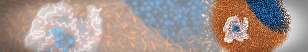
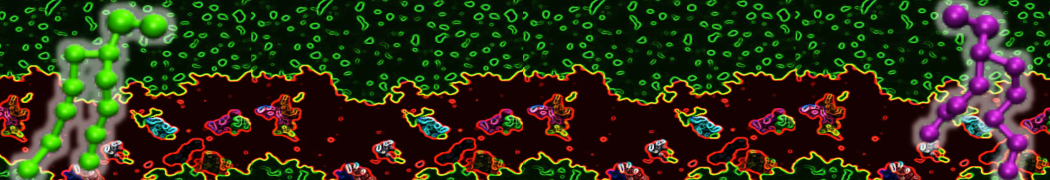
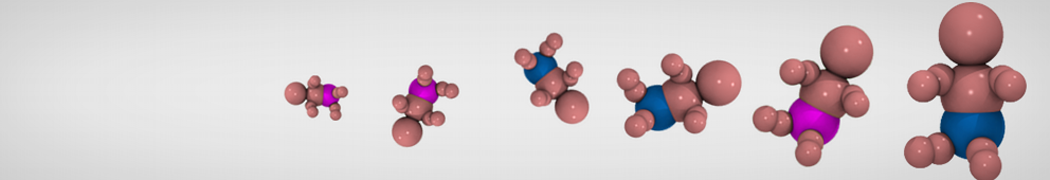
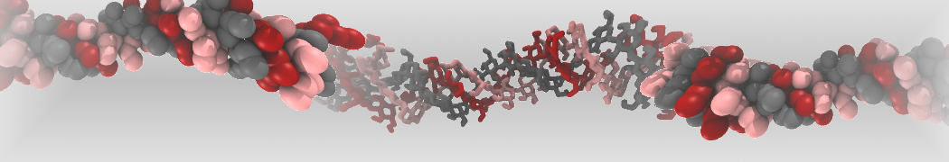
"Effects of PEGylation on the Size and Internal Structure of Dendrimers: Self-Penetration of Long PEG Chains into the Dendrimer Core" H. Lee and R.G. Larson, Macromolecules, ASAP, 2011.

G4 PAMAM dendrimers grafted with poly(ethylene glycol) (PEG) of different sizes (Mw = 550 and 5000) and grafting densities (12−94% of surface terminals) were simulated using the coarse-grained (CG) force fields previously developed and reparametrized in this work. Simulations are carried out for G4, G5, and G7 un-PEGylated dendrimers that are either unprotonated, terminally protonated, or protonated on both terminals and interior sites, corresponding to pH values of >10, 7, and <5, respectively. As protonation increases, simulations show only a small (6% for G4 and G5) change of dendrimer radius of gyration Rg and show a structural transition from dense-core to dense-shell structure, both of which are in agreement with recent scattering experiments and all-atom simulations. For the PEGylated dendrimers, the Rg of the fully PEG(Mw = 5000)-grafted dendrimer also agrees well with experiment. Longer PEG chains with higher grafting density yield PEG−PEG crowding, which stretches dendrimer terminals toward water more strongly, leading to larger size and a dense-shell structure of the dendrimer. Long PEG chains at high grafting densities also penetrate into the dendrimer core, while short ones do not, which might help explain the reduced encapsulation of hydrophobic compounds seen experimentally in dendrimers that are 75%-grafted with long PEG’s (Mw = 5000). This reduced encapsulation for dendrimers with long grafted PEG’s has previously been attributed to PEG-induced dendrimer aggregation, but this explanation is not consistent with our previous simulations which showed no aggregation even with long PEG’s but is consistent with the new simulations reported here that show PEG penetration into the core of the dendrimer to which the PEG is attached.

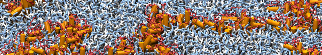
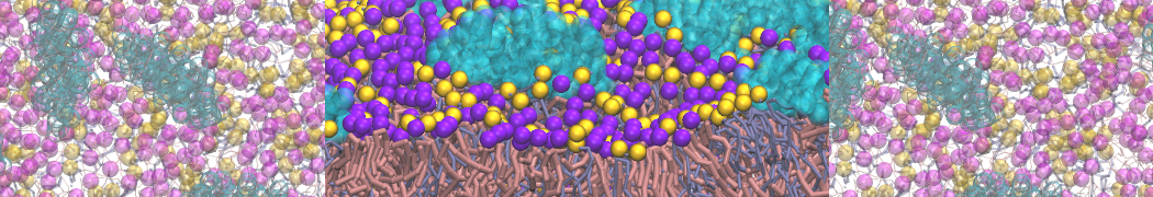
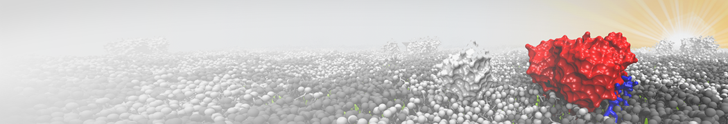
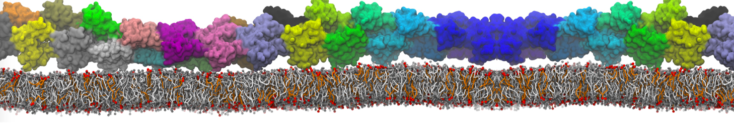
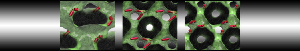
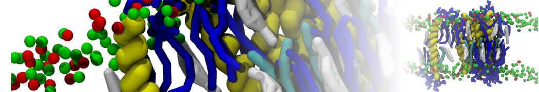
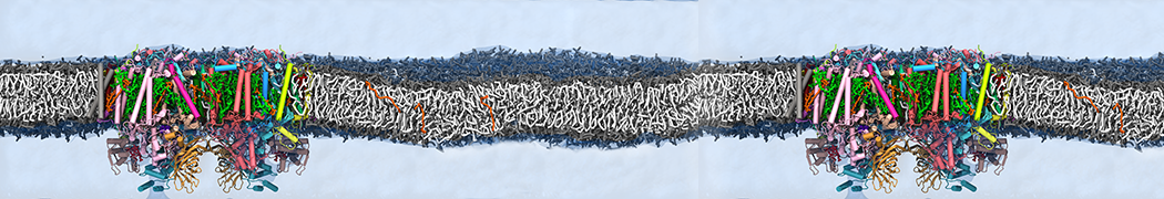
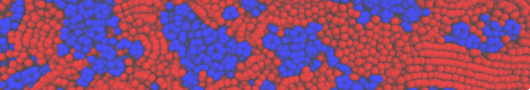
"Membrane Pore Formation Induced by Acetylated and Polyethylene Glycol-Conjugated Polyamidoamine Dendrimers" H. Lee and R.G. Larson, J. Phys. Chem. C, ASAP, 2011.

We performed molecular dynamics (MD) simulations of 36 copies of unmodified (charged), acetylated, and polyethylene glycol (PEG)-conjugated G4 dendrimers in dimyristoylphosphatidylcholine (DMPC) bilayers with explicit water using coarse-grained (CG) lipid and PEG force fields (FF). Attachment of small PEG chains to the dendrimer leads to the same reduction in membrane permeability as does attachment of acetyl groups, while a larger PEG size or a higher degree of PEGylation induces even fewer pores. This indicates that PEGylation is more efficient than acetylation in reducing membrane permeability and cytotoxicity, in qualitative agreement with experimental findings (Kim et al. Bioconjugate Chem. 2008, 19, 1660). Attachment of larger PEG chains makes the dendrimer−PEG complex larger and more spherical. Although a larger size and a more spherical shape are usually conducive to pore formation, a thick PEG layer on the dendrimer surface blocks the charge interaction between cationic dendrimer terminals and anionic lipid phosphate groups, and thus inhibits pore formation, despite the increased dendrimer size. Large PEG chains also keep the dendrimer−PEG complexes far from each other, suppressing interparticle aggregation.