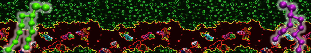
Graphene nanosheets


Dan Wu and Xiaoning Yang, Coarse-Grained Molecular Simulation of Self-Assembly for Nonionic Surfactants on Graphene Nanostructures. J. Phys. Chem. B, Article ASAP. DOI: 10.1021/jp3043939

Self-assembly of amphiphilic molecules on the surfaces of nanoscale materials has an important application in a variety of nanotechnology. Here, we report a coarse-grained molecular dynamics simulation on the structure and morphology of the nonionic surfactant, n-alkyl poly(ethylene oxide) (PEO), adsorbed on planar graphene nanostructures. The effects of concentration, surfactant structure, and size of graphene sheet are explored. Because of the finite dimension effect, various morphological hemimicelles can be formed on nanoscale graphene surfaces, which is somewhat different from the self-assembly structures on infinite carbon surfaces. The aggregate morphology is highly dependent on the concentration, the chain lengths, and the size of graphene nanosheets. For the nonionic surfactant, the PEO headgroups show strong dispersion interaction with the carbon surface, leading to a side edge adsorption behavior. This simulation provides insight into the supramolecular self-assembly nanostructures and the adsorption mechanism for the nonionic surfactants aggregated on graphene nanostructures, which could be exploited to guide fabrication of graphene-based nanocomposites.

Influenza fusion


H.J. Risselada, G. Marelli, M. Fuhrmans, Y.G. Smirnova, H. Grubmüller, S.J. Marrink, M. Muller. Line-tension controlled mechanism for influenza fusion. PLoS ONE 7:e38302, 2012. open access

Our molecular simulations reveal that wild-type influenza fusion peptides are able to stabilize a highly fusogenic pre-fusion structure, i.e. a peptide bundle formed by four or more trans-membrane arranged fusion peptides. We rationalize that the lipid rim around such bundle has a non-vanishing rim energy (line-tension), which is essential to (i) stabilize the initial contact point between the fusing bilayers, i.e. the stalk, and (ii) drive its subsequent evolution. Such line-tension controlled fusion event does not proceed along the hypothesized standard stalk-hemifusion pathway. In modeled influenza fusion, single point mutations in the influenza fusion peptide either completely inhibit fusion (mutants G1V and W14A) or, intriguingly, specifically arrest fusion at a hemifusion state (mutant G1S). Our simulations demonstrate that, within a line-tension controlled fusion mechanism, these known point mutations either completely inhibit fusion by impairing the peptide’s ability to stabilize the required peptide bundle (G1V and W14A) or stabilize a persistent bundle that leads to a kinetically trapped hemifusion state (G1S). In addition, our results further suggest that the recently discovered leaky fusion mutant G13A, which is known to facilitate a pronounced leakage of the target membrane prior to lipid mixing, reduces the membrane integrity by forming a ‘super’ bundle. Our simulations offer a new interpretation for a number of experimentally observed features of the fusion reaction mediated by the prototypical fusion protein, influenza hemagglutinin, and might bring new insights into mechanisms of other viral fusion reactions.


Ganglioside domains


D.H. de Jong, C.A. Lopez, S.J. Marrink. Molecular view on protein sorting into liquid-ordered membrane domains mediated by gangliosides and lipid anchors. Farad. Discuss., accepted. . DOI:10.1039/C2FD20086D

We present results from coarse grain molecular dynamics simulations of mixed model membranes consisting of saturated and unsaturated lipids together with cholesterol, in which lipid-anchored membrane proteins are embedded. Membrane proteins studied are the peripherally bound H-Ras, N-Ras, and Hedgehog, and the transmembrane peptides WALP and LAT. We provide a molecular view on how the presence and nature of these lipid anchors affects partitioning of the proteins between liquid-ordered and liquid-disordered domains. In addition, we probed the role of the ganglioside lipid GM1 on the protein sorting, showing formation of GM1-protein nano- domains that act as shuttles between the differently ordered membrane regions.


Ras nanoclusters

Lorant Janosi, Zhenlong Li, John F. Hancock, and Alemayehu A. Gorfe. Organization, dynamics, and segregation of Ras nanoclusters in membrane domains PNAS  109, 8097, 2012. doi:10.1073/pnas.1200773109

Recent experiments have shown that membrane-bound Ras proteins form transient, nanoscale signaling platforms that play a crucial role in high-fidelity signal transmission. However, a detailed characterization of these dynamic proteolipid substructures by high-resolution experimental techniques remains elusive. Here we use extensive semiatomic simulations to reveal the molecular basis for the formation and domain-specific distribution of Ras nanoclusters. As model systems, we chose the triply lipidated membrane targeting motif of H-ras (tH) and a large bilayer made up of di16∶0-PC (DPPC), di18∶2-PC (DLiPC), and cholesterol. We found that 4–10 tH molecules assemble into clusters that undergo molecular exchange in the sub-μs to μs time scale, depending on the simulation temperature and hence the stability of lipid domains. Driven by the opposite preference of tH palmitoyls and farnesyl for ordered and disordered membrane domains, clustered tH molecules segregate to the boundary of lipid domains. Additionally, a systematic analysis of depalmitoylated and defarnesylated tH variants allowed us to decipher the role of individual lipid modifications in domain-specific nanocluster localization and thereby explain why homologous Ras isoforms form nonoverlapping nanoclusters. Moreover, the localization of tH nanoclusters at domain boundaries resulted in a significantly lower line tension and increased membrane curvature. Taken together, these results provide a unique mechanistic insight into how protein assembly promoted by lipid-modification modulates bilayer shape to generate functional signaling platforms.


Membrane tethers

S. Baoukina, S.J. Marrink, D.P. Tieleman. Molecular structure of membrane tethers. Biophys. J., 102:1866-1871, 2012. open access

Membrane tethers are nanotubes formed by a lipid bilayer. They play important functional roles in cell biology and provide an experimental window on lipid properties. Tethers have been studied extensively in experiments and described by theoretical models, but their molecular structure remains unknown due to their small diameters and dynamic nature. We used molecular dynamics simulations to obtain molecular-level insight into tether formation. Tethers were pulled from single-component lipid bilayers by application of an external force to a lipid patch along the bilayer normal or by lateral compression of a confined bilayer. Tether development under external force proceeded by viscoelastic protrusion followed by viscous lipid flow. Weak forces below a threshold value produced only a protrusion. Larger forces led to a crossover to tether elongation, which was linear at a constant force. Under lateral compression, tethers formed from undulations of unrestrained bilayer area. We characterized in detail the tether structure and its formation process, and obtained the material properties of the membrane. To our knowledge, these results provide the first molecular view of membrane tethers.


Lipid flip-flop

Fumiko Ogushia, Reiko Ishitsukab, Toshihide Kobayashib, Yuji Sugita. Rapid flip-flop motions of diacylglycerol and ceramide in phospholipid bilayers. Chem. Phys. Lett. 522:96–102, 2012

We have investigated flip-flop motions of diacylglycerol and ceramide in phospholipid bilayers using coarse-grained molecular dynamics simulations. In the simulations, flip-flop motions of diacylglycerol and ceramide in the DAPC membrane are slower than cholesterol. Rates correlate with the number of unsaturated bonds in the membrane phospholipids and hence with fluidity of membranes. These findings qualitatively agree with corresponding experimental data. Statistical analysis of the trajectories suggests that flip-flop can be approximated as a Poisson process. The rate of the transverse movement is influenced by depth of the polar head group in the membrane and extent of interaction with water.